| Calculation of Corneal Power | All Features | Upgrades | Technical |
The Pentacam® is a combined device consisting of a slit illumination system and a Scheimpflug camera which rotates around the eye.
A thin layer within the eye is illuminated through the slit. Being not entirely transparent the cells scatter the slit light. In doing so they create a sectional image which is then photographed in side view by a camera. This camera is oriented according to the Scheimpflug principle, thus creating an image of the illuminated plane which appears completely sharp from the anterior surface of the cornea right up to the posterior surface of the crystalline lens. Swivelling around the eye, the slit-camera device generates a series of radially oriented images of the anterior eye chamber. In the subsequent analysis of the sectional images, tissue boundaries are detected and point clouds are assigned to the various tissue layers (anterior and posterior corneal surfaces, iris, crystalline lens).
The sectional images are saved, corrected in relation to a common reference point and then put together to create a three-dimensional model of the entire anterior eye chamber. This makes it possible to generate reproducible tomographic images of the anterior eye chamber in any desired plane.
After correction for Scheimpflug distortion and light refraction at tissue interfaces the exact of location of image edge points in the eye is determined by means of raytracing. Eye movements during image acquisition are captured by a second camera (pupil camera) and also taken into account in the mathematical evaluation. This produces a set of three-dimensional measurement data which gives a precise geometric description of the anterior eye segment. This data in turn can be used to generate data on elevation, curvature, pachymetry, depth of the anterior eye chamber…etc. in the well-known form of colour maps.
The Pentacam® is the only instrument on the market able to perform a precise and complete measurement and analysis of the centre of the cornea. The rotating measurement principle avoids measurement errors that would result from horizontal scanning. Due to the radial orientation of the sectional images the density of data points is greatest at the centre.
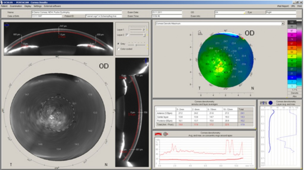
Corneal Optical Densitometry
The Pentacam® / Pentacam® HR software allows customized and objective standardized evaluation of corneal optical densitometry. It is displayed as a color coded map over its full depth and area and can be assessed individually in different layers and zones. The respective layer depth and zone size are displayed in the two Scheimpflug images as well as in a colored or a grey scaled map.
For study and normative data analysis it is displayed in fixed layers and zones in the table chart. The objective analysis of corneal optical density allows the detection of corneal Fuchs dystrophy or corneal scars for example. It provides an objective follow-up before and after CXL, PRK, LASIK, DSEK, etc. as well.
Corneal Ring Display
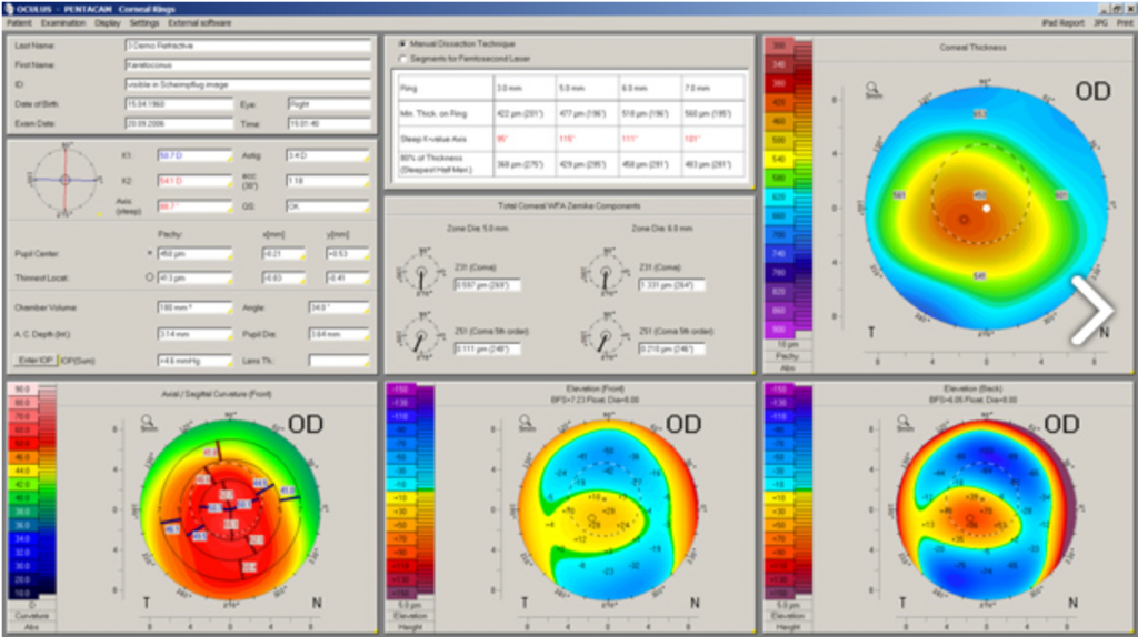
The pre-op planning for implanting a corneal ring is a combination of several parameters. The Corneal Rings
Display includes all relevant information for this purpose.
The table chart in the upper center contains the minimum thickness for manual dissection technique or using a Femto laser to design the channel.
Below the table chart, the 3rd and the 5th order coma amount and axis are shown which together with the astigmatism axis helps orientating the ring(s). The colored maps are helpful for the qualitative inspection of the corneal properties.
The corneal ring display in combination with the link to the external Ferrara Ring calculator software realizes an automated and comprehensive clinical use. The Ferrara Ring calculator imports all necessary data from the Pentacam® and suggests the best ring type and orientation axis.
Please keep in mind that the mentioned external software might not be available in your country.
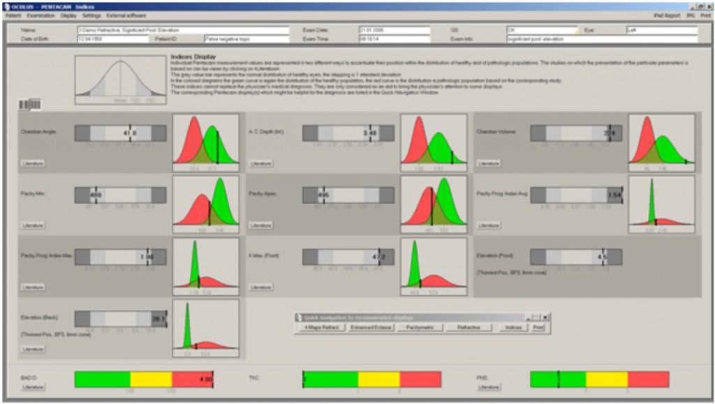
Indices Report
The Indices Report allows a quick screening of new patients to detect abnormalities. The intuitive guide recommends further displays to look at.
Important parameters of the anterior eye segment, displayed with the Pentacam® / Pentacam® HR software, are analyzed and validated in published papers and articles.
Their normal (green) and pathological (red) distribution is displayed in diagrams.
This presentation provides an intuitive, quick but comprehensive overview.
For detailed assessment, the literature source used is also mentioned. Based on individual results, the navigation bar recommends further displays to review to get additional information.