
The OCULUS Easyfield® C is designed for use as a visual field screener and a threshold perimeter for immediate re-examination of any abnormal findings. Full compliance with Goldman Standards makes the Easyfield® C ideal for all common examinations of the cent
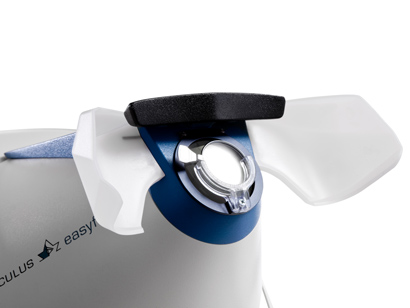
ral field of vision up to 30°. The Easyfield® C has an adjustable double chin rest and eye shields for maximum patient comfort.
Request A Quote | Request A Free Demo
Ergonomic Design
One of the most striking features of the Easyfield® C is its small space requirement. The self-contained design and light-shielded viewer enable examinations to be conducted in normal room lighting conditions. Its robustness and low weight make the Easyfield® C Perimeter ideal for portable use. The lack of moving device parts guarantees long product life
Easy Operation
The Easyfield® C Perimeter can be controlled via an external computer (Notebook or PC). The familiar user interface of the OCULUS programs is available on the computer and you can enjoy the full freedom of networking the examination data. The use of translucent Easyfield® C occluders allows you to conduct examinations without the usual eye patch, thus saving you valuable time as you prepare for the examination.
Increased Diagnostic Reliability
Additional software programs improve the diagnostic value of visual field examinations conducted with the Easyfield® C Perimeter. The Glaucoma Staging System (GSS2)per Dr. Brusini and the Glaucoma Staging Program (GSP) are Glaucoma risk indicators. The Threshold Noiseless Trend (TNT) is used for efficient progression analysis.
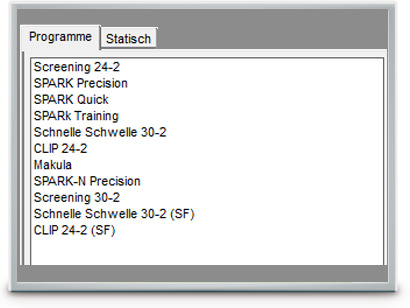
The Examination Programs
The Easyfield® C provides pre-defined programs for the examination routines that are most frequently used. Measurements of the central visual field or the macula area are thereby conducted. This program series can be extended, if necessary, by interlinking available test grids and test strategies.
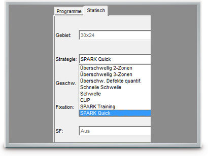
The OCULUS Test Strategies
As a screening unit, the Easyfield® C Perimeter generally uses threshold related suprathreshold examination
strategies. The advantage of this is that despite the short examination time, the examiner gains a meaningful overview of the examined area. Multiple test strategies are available for determination of the exact numeric values of
the perception thresholds. (We prefer not to promote CLIP anymore.) The OCULUS “Fast Threshold” achieves a similar performance with clever improvements to the classic 4-2 step method. The unique SPARK strategy provides a rapid and reproducible threshold measurement for glaucoma patients.
The OCULUS Test Point Grid
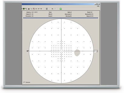
The Easyfield® C Perimeter’s fixed light emitting diode grid permits examination in areas such as the 30-2, 24-2 or 10-2. It is also possible to test individual hemispheres or quadrants. If necessary, individual points can be examined in any combination. The modular setup of the test programs allows all areas to be examined using all available standard strategies.
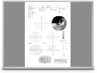
Results Printout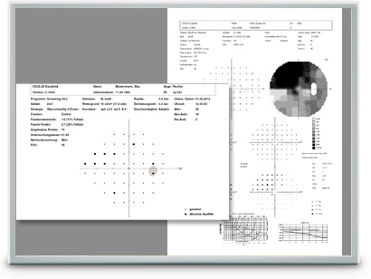
The measuring results of the Easyfield® C Perimeter are summarized in a standard printout. For suprathreshold tests, only a single informational overview is printed, whereas for threshold examinations, all clinically relevant data are recorded and shown in various depictions.
SPARK Strategy
The SPARK examination strategy was primarily developed for Glaucoma patients and is available for all OCULUS perimeters. It is based on data from more than 90,000 perimetric diagnoses and allows a very fast, yet very precise measurement of the thresholds in the central field of vision. The modular configuration of the method makes it suitable for a variety of applications:
- SPARK Precision (Optional software) is the full version. The complete examination of the field of vision of glaucoma patients takes only 3 minutes per eye. The greater stability of the results allows for much better progression analysis.
- SPARK Quick is the strategy used for progression or screening examinations. The test takes just 1.5 minutes per eye.
- SPARK Training is ideal for patient training. The 40 second measurement can also be used as a screening examination.
Glaucoma Staging System (GSS 2) per Brusini
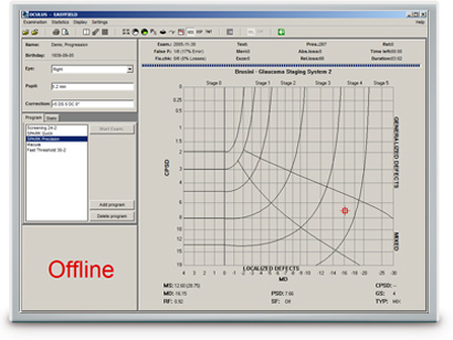
The Glaucoma Staging System (GSS 2) per Brusini classifies visual field defects based on the perimetric indices MD (Mean Deviation) and PSD (Pattern Standard Deviation). The representative point of the examination is placed in a chart in accordance with the values of these indices. The chart shows defined areas for the different stages of disease (Stage 0 – Stage 5). It also separates generalized, localized and mixed defects.
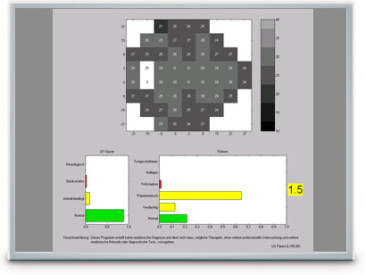
Glaucoma Staging Program (GSP)
The Glaucoma Staging Program (GSP) is useful in early detection of Glaucoma.
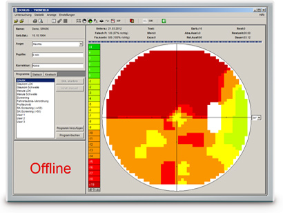
The GSP software grades examination findings into visual field classes (normal, glaucomatous, artifactual and neuro) based solely on their appearance. In addition, risk classes (normal, suspect, pre-perimetric, early stage, moderate and severe) are also assigned to findings of different severities that are classified as normal or glaucomatous. The examination results are presented in easy to understand green, yellow and red bar charts.
The striking novelty of the GSP is its ability to detect subtle changes in the visual field associated with early stage Glaucoma. Findings in the suspect and pre-perimetric risk classes contain reductions in the visual field that cannot be readily seen by the examiner. They usually remain undetected by the standard perimetric indices.
The Glaucoma Likelihood Index (GLI) summarizes the results of the GSP classification into a single parameter, presenting a value of between 0 (normal) and 5 (advanced Glaucoma).
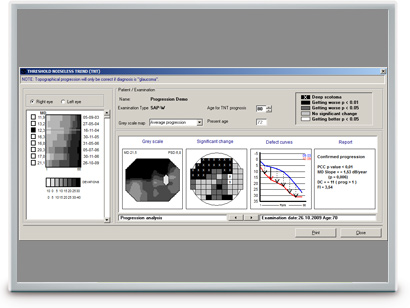
Threshold Noiseless Trend (TNT) Progression Analysis
TNT provides a quantitative, statistical analysis of the visual field examination results conducted over time. For follow-up purposes, the software uses the visual field results and patient’s threshold values supplied by 30-2, 30×24 or 24-2 grids over the entire examination period. TNT uses a specific filter to reduce the fluctuation range of the threshold values and to perform a consistent trend analysis. In conjunction with the fast SPARK strategy, the sensitivity of detecting progression of early stage glaucoma is greatly improved.
- TNT creates a concise progression analysis report containing the most important parameters (MD increase, p-values, etc.).
- TNT can differentiate between diffuse and focal progression based on the focusing index (FI) value.
- TNT applies multiple statistical criteria to establish possible progression.
- TNT displays the prognosis of the expected visual field for a patient age-group selected by the examiner.
| Programs: | Pre-defined glaucoma, macula, screening and neurological tests User-defined tests |
| Test patterns: | 30-2, 24-2, 30×24, 10-2, hemisphere, customized patterns |
| Strategies: | Threshold strategies: SPARK Quick, CLIP, OCULUS Fast Threshold, Full Threshold (4/2) Optional: SPARK Precision Age adapted suprathreshold screening (2-zone, 3-zone, quantify defects) |
| Examination Speed: | Adaptive, fast, normal, slow, user-defined |
| Fixation control: | CMOS camera, through central threshold, Heijl-Krakau (using the blind spot) |
| Result Display: | Glaucoma Staging System (GSS2), Glaucoma Staging Program (GSP) |
| Reports: | Threshold Noiseless Trend (TNT) progression report |
| Perimeter bowl radius: | r = 30 cm (11.8 in) |
| Maximum eccentricity: | 30° |
| Stimulus size: | Goldmann III |
| Stimulus luminance range / increments: | 0.03 – 3,180 cd/m² (0.1 – 10,000 asb) / 0.1 log steps |
| Background luminance: | 10 cd/m² (31.4 asb) |
| Stimulus color: | White |
| Stimulus duration: | 200ms / user-defined |
| Patient positioning: | Measurement head with adjustable angle of inclination, adaptable double chinrest, double headrest |
| Software: | Device control, patient management, backup, and print software (Windows®) Built-in networking, easy EMR-integration, DICOM compatibility |
| Interface: | USB, RS232 |
| Dimensions (W x D x H) | 316 x 506 x 435 mm (12.4 x 19.9 x 17.1 in) |
| Weight with accessories | 6.8 kg (15.0 lbs) |
| Max. power consumption | 30 W |
| Voltage and frequency |
100/240 V AC 50-60Hz |
| Min. computer requirements | Operating system: Windows® XP |

![]()
![]() OCULUS is certified in acc. with
OCULUS is certified in acc. with
DIN EN ISO 13485