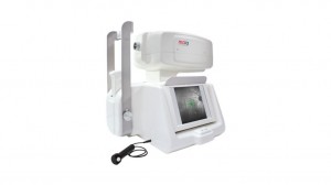
Overview
MAIA is a new generation fundus perimeter that helps monitor the course of retinal diseases and the efficacy of treatment. It measures macular sensitivity, fixation, stability, and the locus of fixation.By comparing macular indexes in patients with and without retinal pathologies, MAIA is very effective at measuring functional changes due to disease and to treatment.
Benefits of Maia:
· Simple to use, patients can be tested in less than 3 minutes per eye
· Compares measured threshold values with a reference database of normal subjects
· Helps monitoring the course of retinal diseases and the efficacy of treatment
· Effectively combines a linear SLO system and a fundus controlled perimetric exam, providing clear and detailed retinal images
How it works:
maia collects both anatomical and functional data. Maia subjectively analyzes macula photoreceptor sensitivity and patient fixation stability in combination with a linear SLO tracked fundus image
· Live imaging of the central retina over a 36° field of view, acquired under IR illumination at 850nm, using a confocal imaging system
· Eye-tracking, recorded at 25 Hz, throughout the test ensures consistent patient fixation
· Subjective measurements of differential light sensitivity at multiple locations in the macula are obtained as in fundus perimetry.
TECHNICAL SPECIFICATIONS
Fundus Imaging
· Line scanning laser ophthalmoscope
Field of view: 36° x 36°
Digital camera resolution: 1024 x 1024 pixel
Optical resolution on the retina: 25 µm
Optical source: superluminescent diode at 850 nm
Imaging speed: 25 fps
Working distance: 30 mm
Other Features
· Minimum pupil diameter: 2.5 mm
Focus adjustment range: -15D to +10D (auto-focus)
Automatic OD/OS recognition
Autofocus