Topography Made Easy With Oculus Keratograph 5M
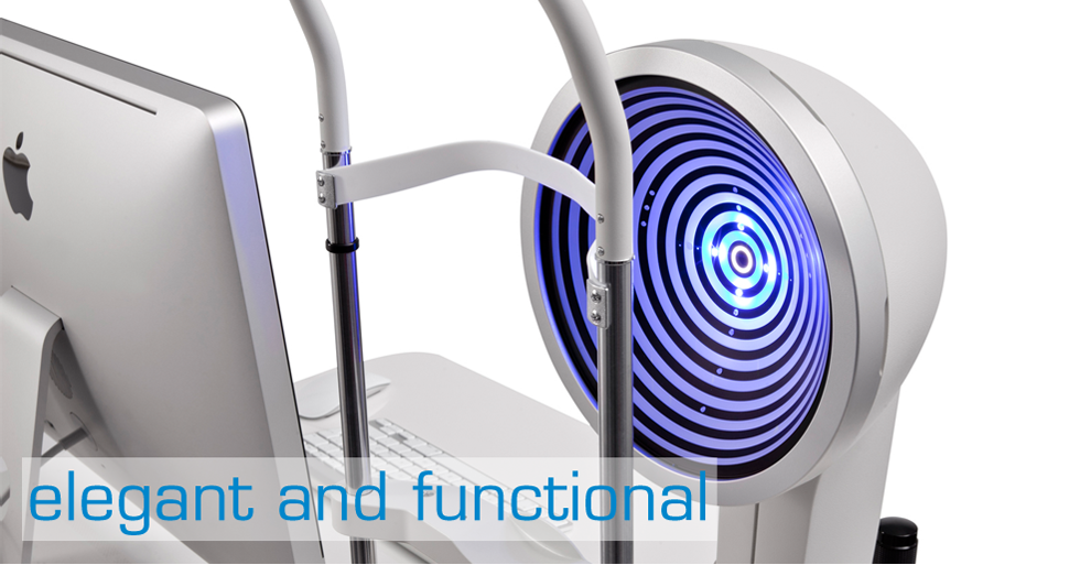
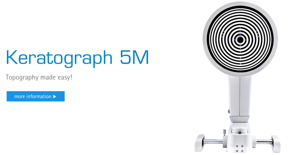
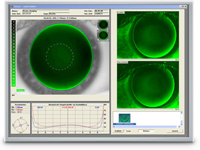
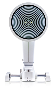
The OCULUS Keratograph 5M is an advanced corneal topographer with a built-in real keratometer and a color camera optimized for external imaging. Unique features include examining the meibomian glands, non-invasive tear film break-up time and the tear meniscus height measurement and evaluating the lipid layer. The high-resolution color camera and the integrated magnification changer offer a new perspective to the tear film assessment procedure.
In addition to these unique features, the Keratograph 5M precisely measures corneal topography. The built-in real Keratometer and the automatic measurement activation guarantee perfect reproducibility of the Ks. The data from the non-contact measurement is automatically analyzed and shown in comprehensive presentation formats.
Gain Trust
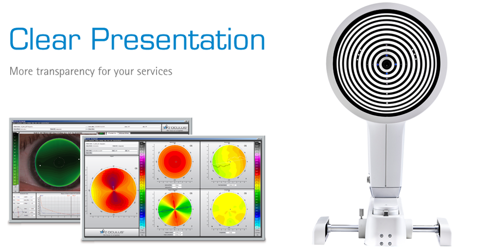
Actively integrate the Keratograph 5M into your practice as a patient consultation tool. With many visual maps and easy to understand diagrams, the Keratograph 5M enhances the communication with your patients. Use your Keratograph 5M as a marketing tool and gain your patients’ confidence.
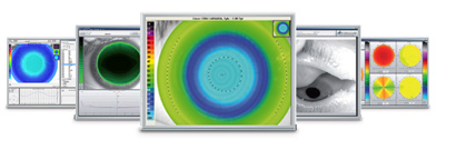
With the Keratograph 5M, you can show your patients images they have never seen before. Gain patient trust by providing professional consultation during examinations and follow-ups.
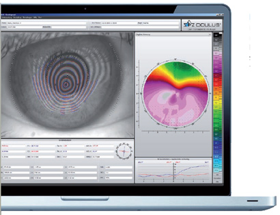 Easy-to-Understand Presentation
Actively integrate the Keratograph into your consultation. Many easy to understand displays support you in communication and patient education. Use your Keratograph 5M as a marketing tool to make your services transparent!
With the TF-Scan software, you can analyze and document the tear film break up time and share the results with your patients.
Let images and videos speak for themselves during your consultation — this creates a strong physician/patient relationship.
Measurements with Placido Ring Illumination
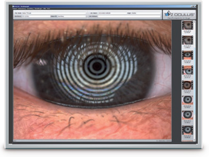
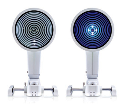
Thousands of measuring points are used to measure the whole surface of the cornea. A white ring illumination is used for this purpose. An infrared ring illumination is also provided for analysis of the tear film to prevent glare-related reflex secretion.
Measurements with Light Emitting Diodes
The perfect illumination has been integrated for every function of the Keratograph 5M: White diodes for the tear film dynamics, blue diodes for fluo-images, infrared diodes for Meibography.
| Accuracy: | ± 0.1 D |
| Reproducibility | ± 0.1 dpt |
| Number of rings: | 22 |
| Working distance: | 80 mm |
| Number of evaluated data points | 22,000 |
| Camera: | Digital CCD camera |
| Light source: | Placido illumination: White Placido illumination: Infrared 880 nm Fluorescein illumination: Blue 465 nm Meibography: Infrared 890 nm Tear film dynamics: White Pupillometer illumination: Infrared 880 nm |
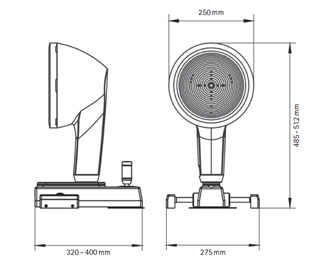
| Dimensions (W x D x H): | 275 x 320 – 400 mm x 485 – 512 mm |
| Weight | 6.8 kg |
| Minimum PC requirements | Processor: Intel Core i3 or better, 4GB RAM, Hard drive space: min. 500 GB, Graphic card: Intel HD Graphics 2000 or better, recommended screen resolution: 1920 x 1080 (Full-HD) |
TF-Scan
The tear film is assessed using either white or infrared illumination. The new high-resolution color camera makes even the finest of structures visible. In addition to NIKBUT (Non-Invasive Keratograph Break-Up Time) and measurement of the meniscus tear, the TF-Scan can also make an assessment of the lipid layer and the tear film dynamics. Tear film analysis with the OCULUS Keratograph 5 M is non-invasive and is conducted without any additional tools.
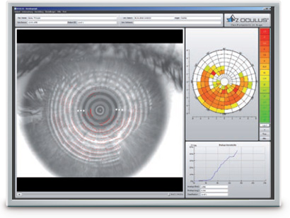
NIKBUT
Assessment of the Tear Film Break-Up Time
The tear film break-up time is measured non-invasively and fully automatically. The new infrared illumination is not visible to the human eye. This prevents glare during the examination. The TF-scan presents the results in a way that is easy for you and your customers to understand.
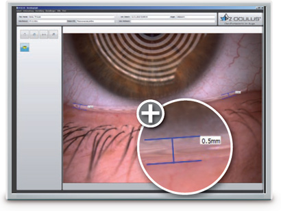
Meniscus Tear Height Assessment of the Tear Film Quantity
The height of the torn meniscus can be precisely measured with an integrated ruler and various magnification
options, and its development along the edge of the bottom lid can be assessed. The results are saved to the patient file.
Lipid Layer
Assessment of the Interference Phenomenon
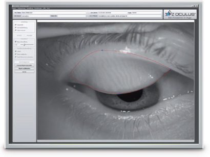
The interference colors of the lipid layer and their structure are made visible and can be recorded. The thickness of the lipid layer is assessed based on the structure and color.
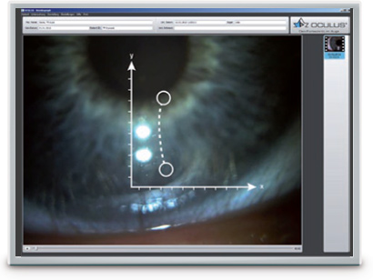
TF Dynamics
Assessment of the Particle Flow
The video recording, with up to 32 frames per second, enables the observation of the tear film particle flow, from which conclusions regarding the viscosity of the tear film can be drawn.
Professional: $19.37
Global: $38.45
Professional: $3.06
Global: $21.43
